Introduction
The immense capacity and specificity of memory storage in the mammalian brain is thought to depend on the plasticity of synaptic connections. Dysfunction of synaptic plasticity is implicated in a range of disorders from Alzheimer’s disease to mental retardation and development of chronic pain states. Understanding how neural activity patterns are translated into lasting changes in synaptic connectivity is therefore one of the most important challenges in basic and clinical neuroscience.
Long-term memory relies on lasting changes in synaptic connectivity, and these synaptic modifications depend on new gene expression and protein synthesis. Arc (activity-regulated cytoskeleton-associated protein), is an immediate-early gene that is rapidly induced upon neuronal activity. Arc transcription, translation, and protein function provides a finely-tuned system for converting neuronal activity patterns into protein synthesis-dependent synaptic plasticity and memory storage. By a biological logic that is not yet understood, Arc synthesis controls both long-term potentiation (LTP) and long-term depression (LTD) of synaptic connections.
Aims
A novel, drug-controllable tool for visualizing new protein synthesis, called TimeSTAMP, is used to visualize and monitor local synthesis and trafficking of Arc in hippocampal neuronal cells undergoing functional changes such as LTP and LTD in the brain of rats. This approach will give us fundamental new insights into how memory operates at the synaptic level.
In the TimeSTAMP reporter, the cassette encoding the protein of interest, e.g. Arc, is followed by a sequence-specific protease, an epitope tag and a fluorescent protein called Venus. The protease in this cassette can be specifically inhibited by the cell permeant drugs. Upon translation of Arc, protease activity cleaves the epitope tag and a part of the Venus fluorescent protein effectively disallowing detection of signal. In the presence of inhibitor the entire fluorescent protein is preserved, so that it can mature and emit fluorescent light upon excitation, thus allowing selective detection of newly synthesized proteins both by fluorescence and epitope tagging.
Methods
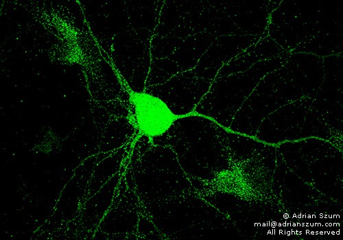
TimeSTAMP allows live time-lapse imaging of new protein synthesis using the fluorescent protein signal in addition to detection of the epitope tagged protein in fixed samples. In this study, in vitro hippocampal neurons dissected out from rat embryos are transfected with this drug-controllable reporter, followed by chemical LTP stimulation and imaging of the temporal and spatial characteristics of Arc by confocal microscopy.
Adrian Szum
Supervisor: Prof. Clive Bramham, Neuroscience